Pelvic Ultrasound
· Bulky size uterus, endometrial lining thickness 13mm, clear endo-myometrial junctional zone, the myometrial layer is heterogenous in texture, no fibroid
· Normal cervix
· Right ovary normal in size, no mature dominant follicle
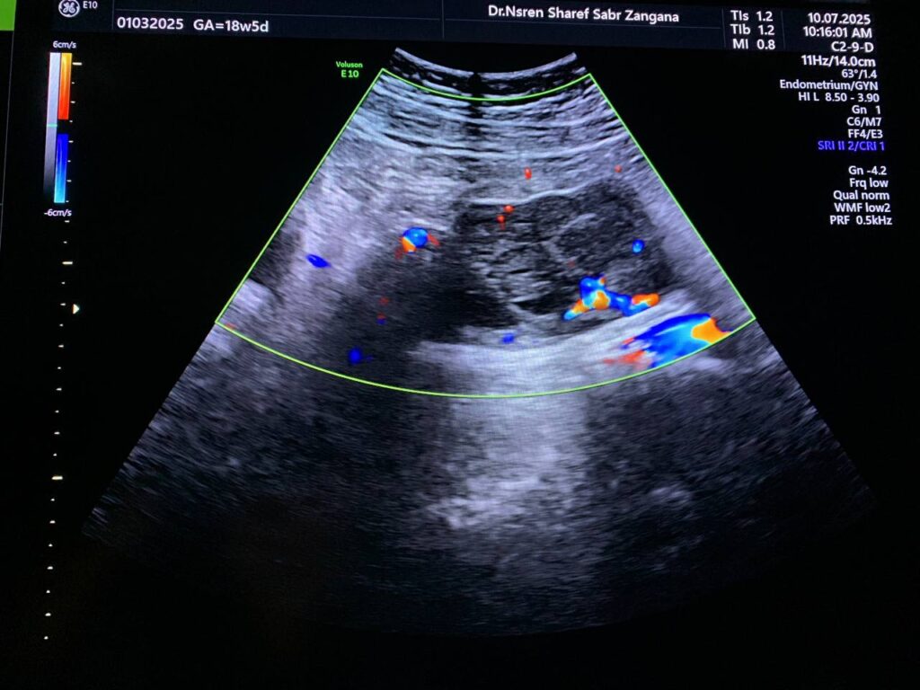
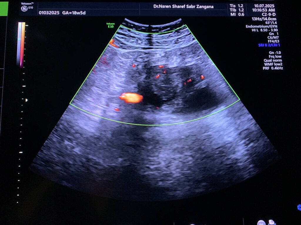
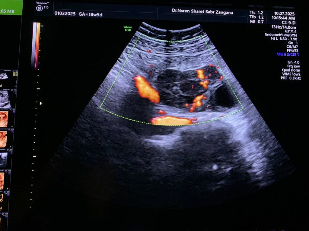
· Left ovarian solid mass vascular on color doppler score 2-3 , its size 57x48mm, no ascites , no enlarged lymph node seen, picture mostly ovarian tumor , please for further study.
· No adnexal mass
· No free pelvic fluid seen

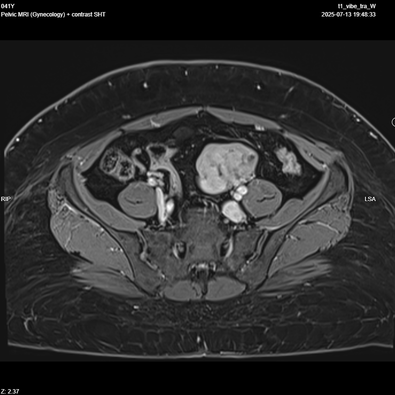
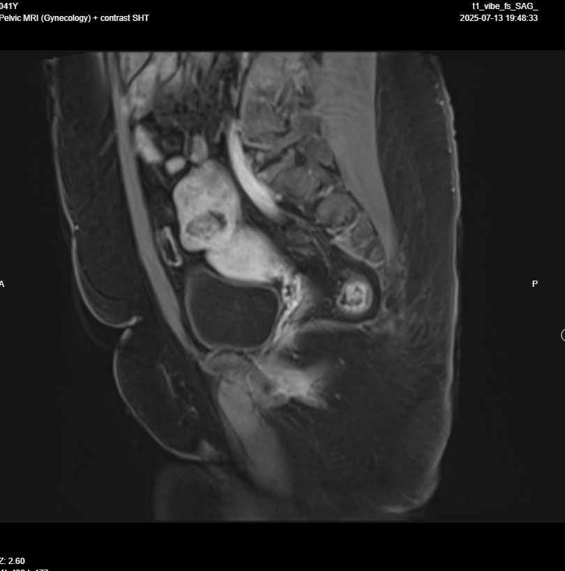
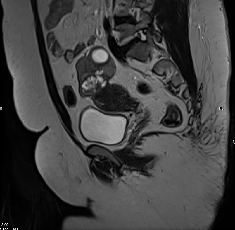
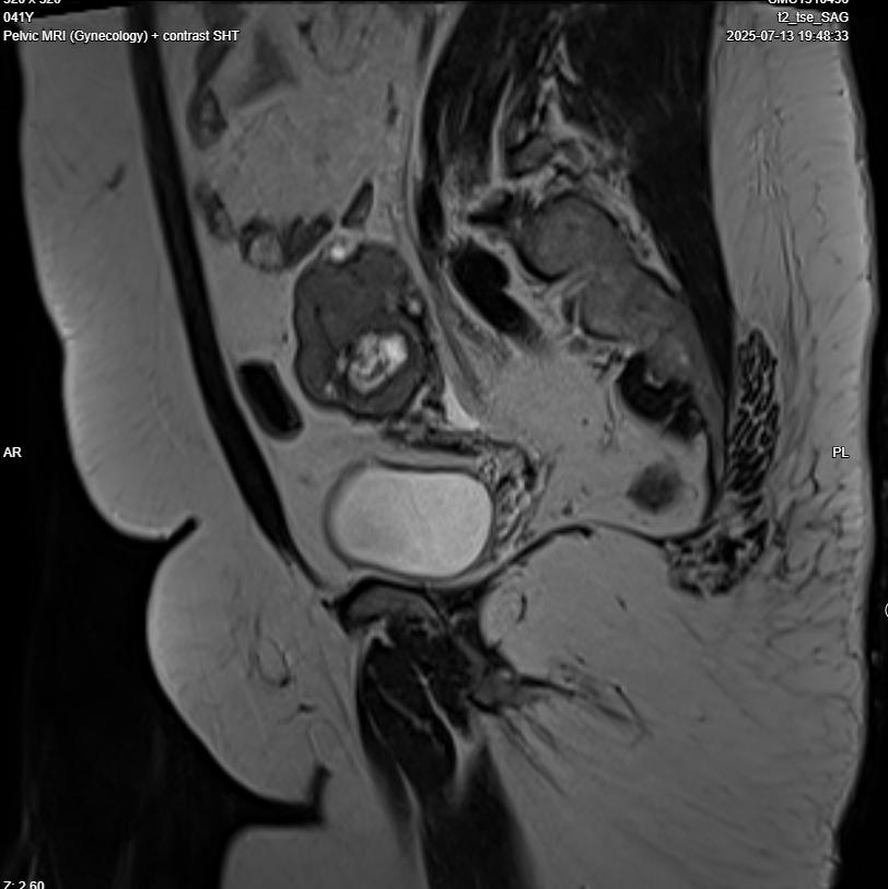
Pelvic MRI with IV contrast
· Evidence of left adnexal irregular lobulated shape solid mass replacing most of left ovarian parenchyma , T1 iso intense , T2 hypo intense , restricted in DWI, no fat , no calcification , after giving contrast showing diffuse progressive hyper enhancement , with high risk curve of enhancement , these radiological features highly suspicious for malignant ovarian tumor, O-RADS 5.