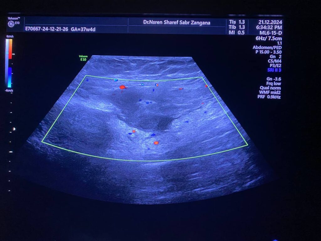
Pelvic Ultrasound
· Normal size uterus, endometrial lining thickness 10mm, contains endometrial polyp about 20x8mm , clear endo-myometrial junctional zone, no fibroid
· Normal cervix
· Right ovary normal in size, dominant follicle 21mm +corpus luteum 14mm
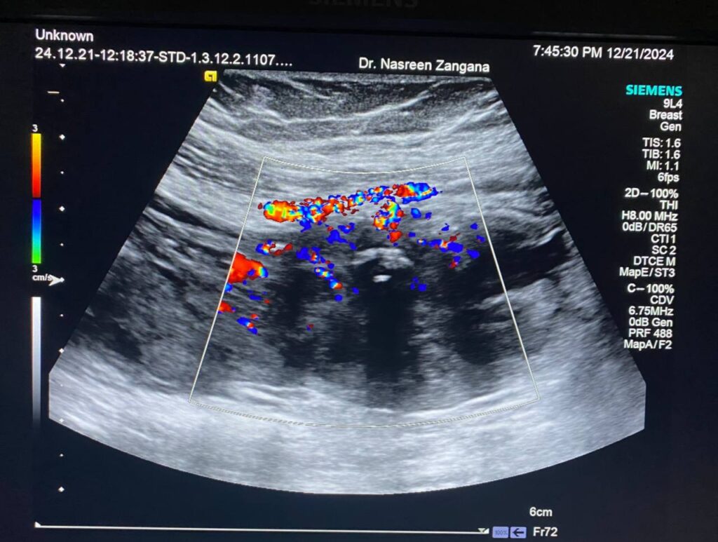
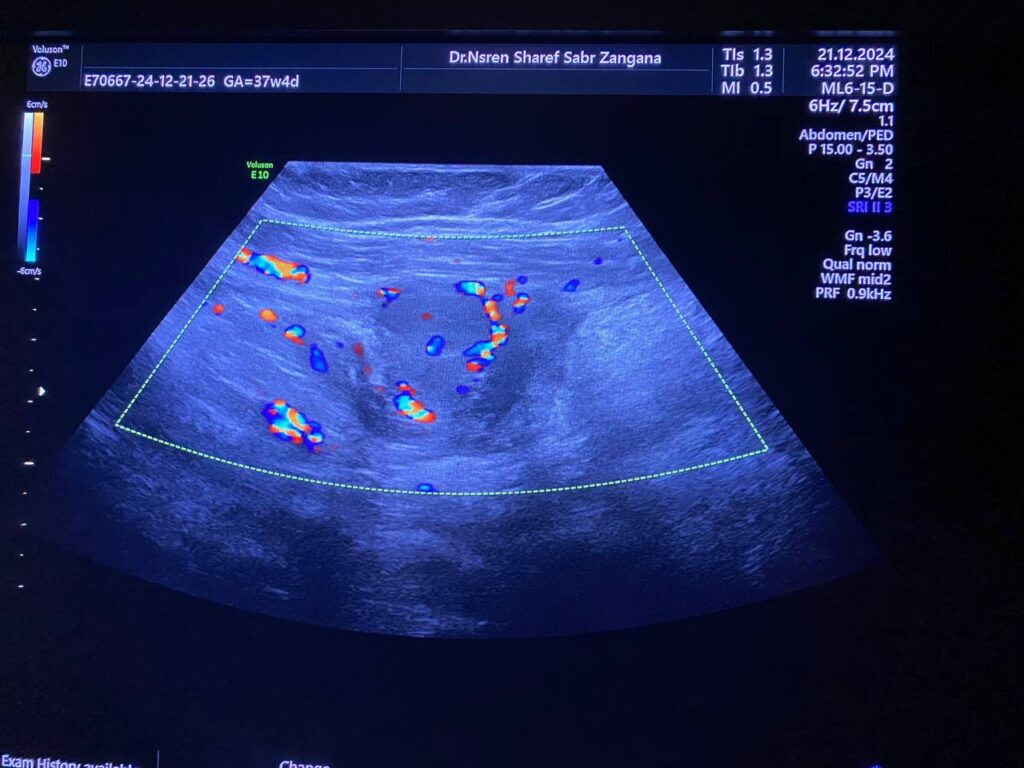
· Left ovary , normal in size , no dominate follicle , with presence of heterogenous vascular solid mass, contain calcification size about 44x30mm with indistinct margin, vascular on color doppler, score 3-4 located just the lateral to the left ovary , with invasion of the adjacent peritoneal & muscular layer ( left rectus abdominis muscle) , mostly suspicious mass , its invasion to the left ovary cannot be excluded , picture could be Desmoid tumor ? peritoneal tumor > , for further study please . no suspicious lymph node seen
· Small amount of free pelvic fluid , could be mild ascites ?? or from corpus luteum of the right ovary ?
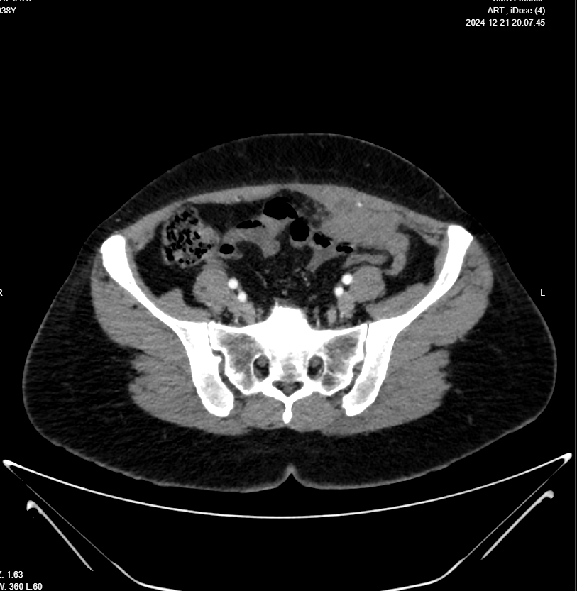
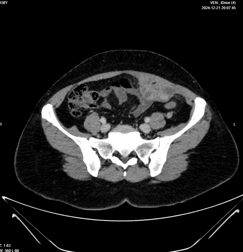
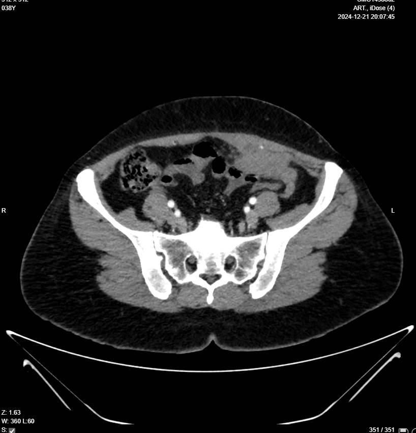
Chest, Abdomen & Pelvic CT With IV Contrast
Clinical data: Malignant LEFT OVARIAN MASS KINDLY FOR PRE RX STAGING
- Clear lung parenchyma, no suspicious parenchymal lesion.
- No pathological hilar or mediastinal LAP, no mass.
- Normal trachea and bronchial tree.
- No pleural or pericardial effusion.
- Evidence of 6 X 4 X 3cm multilocular mass in Lt lower abdomen, arises from Lt adnexa “attached to uterus and Lt ovary”, invades posterior aspect of Lt rectus muscle, in contact with Lt inferior epigastric artery, also in contact with loop of small intestine with no frank invasion, this mass has surrounding fat stranding, which makes it possible to be infection like tubo-ovarian abscess, although malignancy can not be excluded, lab studies and MRI is suggested for further evaluation please.