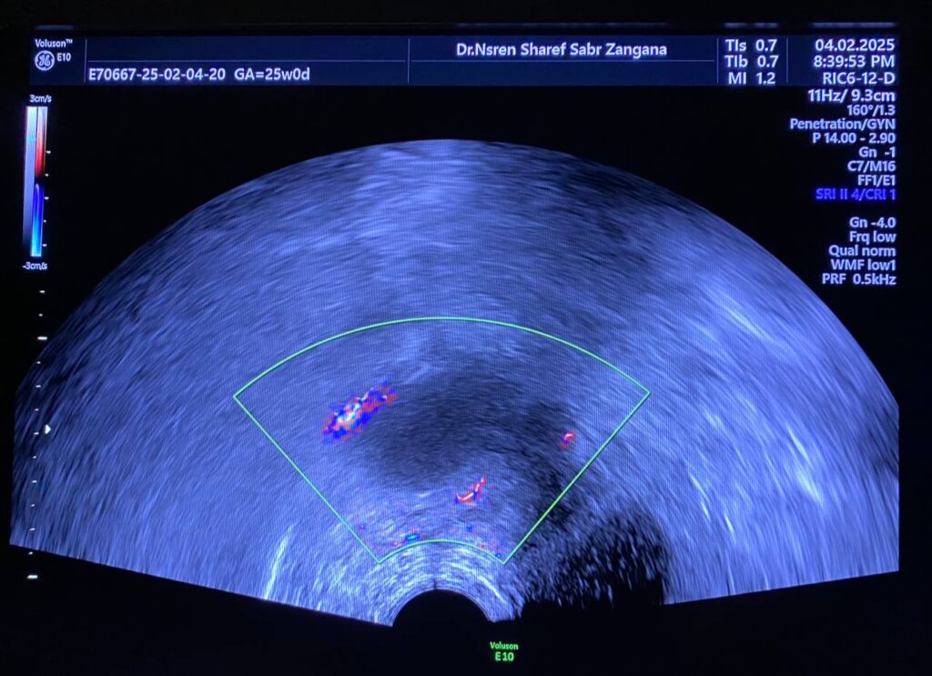
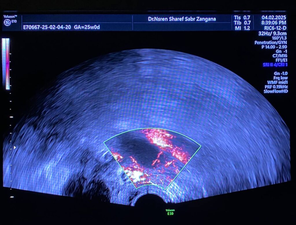
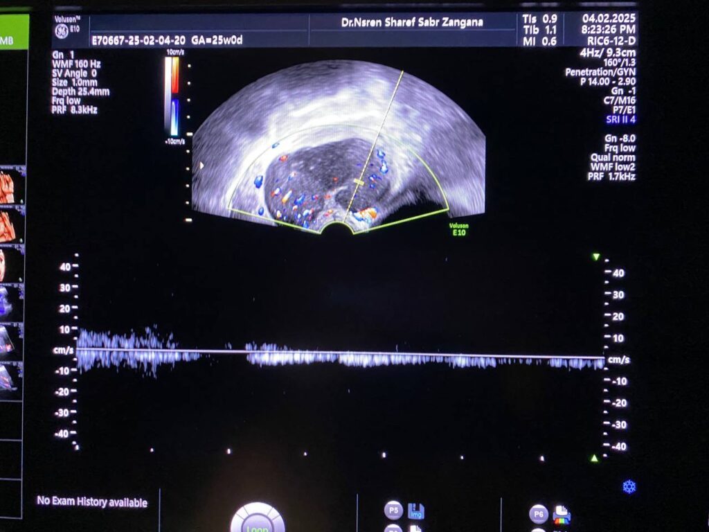
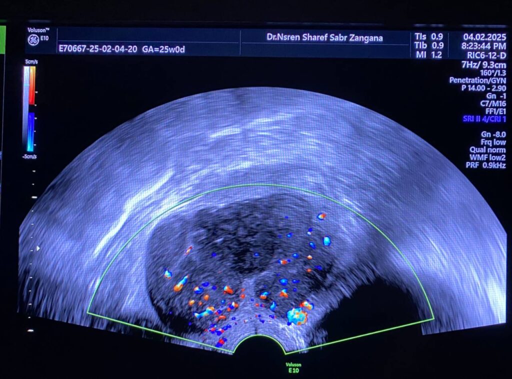
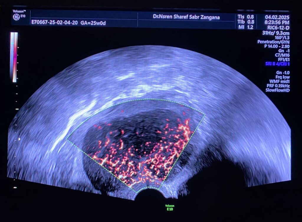
Bulky uterus, endometrial lining thickness 2mm, occupied by clear avascular fluid , mostly due to mass effect , Presence of hypoechoic solid mass 68x48mm ,vascular on color Doppler , Color Score 4 (Hyper vascular) , within right sided posterior submucosal subserosal region , associated with other two smaller masses seen on the anterior wall , their sizes are 19x18mm& 8x12mm (Metastasis? Lymphoma ??) or other suspicious masses
Associated with Presence of pathological lymph node in the right para iliac region size 35x24mm& other pathological lymph node in the left para iliac region 35x21mm , lymphoma or secondary metastasis can not be excluded
Atrophied both ovaries, bilateral few tiny follicles, their sizes less than 2mm, no dominate follicle, nor mass
No free pelvic fluid


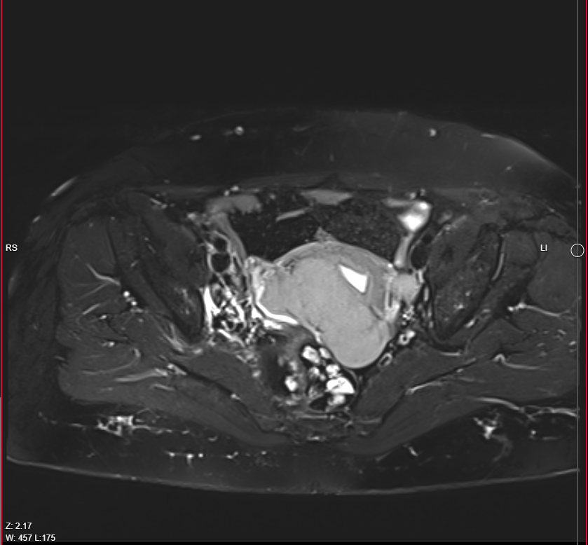
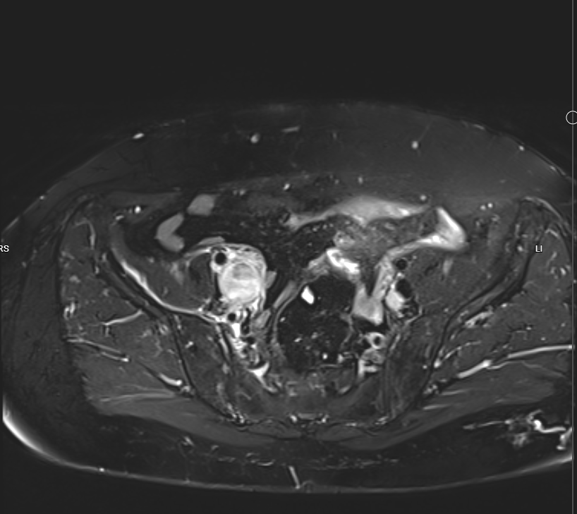
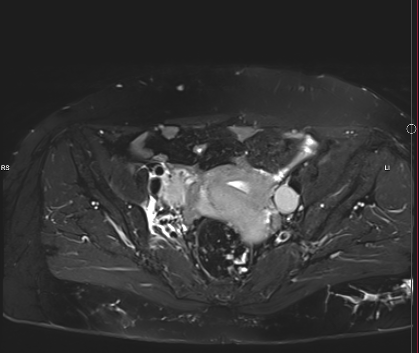
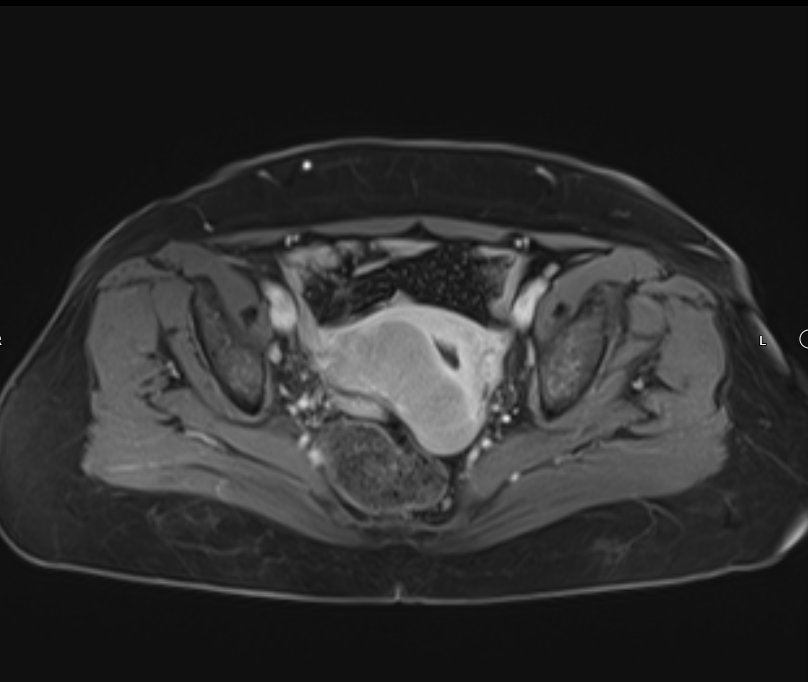
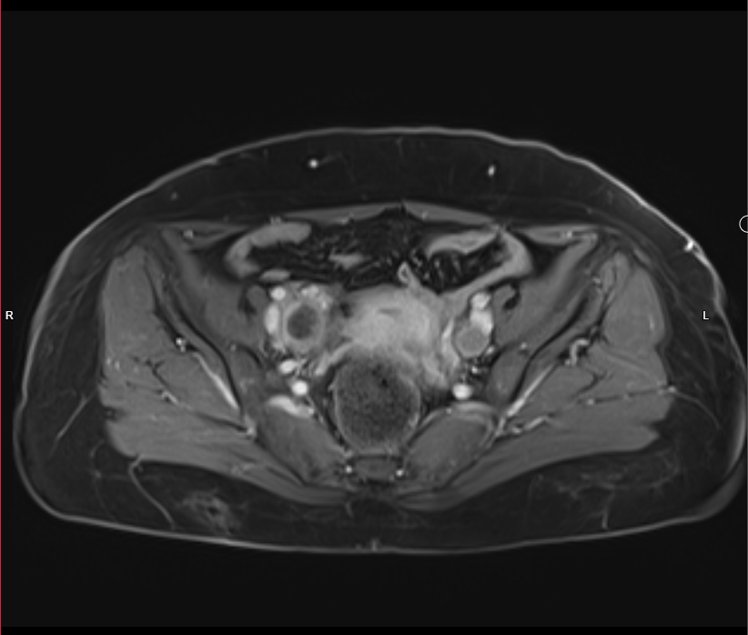
Pelvic MRI With IV Gd:
- Evidence of 7.5 X 4cm mass involves Rt side of uterus reaches endometrium, involves whole thickness of uterus and extends to Rt adnexa, also infiltration of peritoneal reflesion in Rt side of rectouterine pouch, shows marked DWI restriction, picture is suggestive of malignant tumor ?lymphoma ?sarcoma
- Single Lt internal iliac pathological LN, 18mm in short axis diameter, its signal is like uterine mass.
- There is also 26 X 22mm lesion in Rt internal iliac region, but heterogeneous signal ?pathological LN with necrosis
- Endometrial thickness is 8 mm, no obvious focal mass.
- Mild pelvic free fluid.
- Normal both ovaries in size, no mass.
- Normal UB in wall thickness.
- No obvious rectal pathology.
- No pelvic bone lesion.